FGA PhD-student Cijsouw and PI Toonen present a new mouse model, expressing a fluorescently tagged version of a protein, Munc18-1. This protein is essential for synaptic transmission. Their findings are published in the march issue of the JCB.
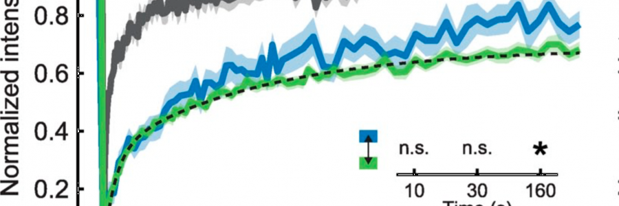
Munc18-1 redistributes in nerve terminals in an activity- and PKC-dependent manner
Synaptic communication
Nerve cells (neurons) communicate with each other via the release of chemical signals at contact sites called synapses. A neuron releases these signals at one side of the synapse and another neuron detects these at the other side. “Much like the words coming from your mouth when you talk and someone else listens with his ear‚, explains first author, Tony Cijsouw.
The ‘mouth’ side of the synapse contains vesicles, filled with neurotransmitter. They fuse with the cell membrane to secrete their content during neuronal activity. To process information, the brain is constantly changing the strength of individual synapses. Deregulation of this process is associated with several brain diseases. “Hence, detailed knowledge of this process is vital to understand neuronal communication and to develop treatments for neurological disorders‚, adds senior author Dr. Ruud Toonen.
New mouse model
Cijsouw and Toonen present a new mouse model, expressing a fluorescently tagged version of a protein, Munc18-1. This protein is essential for synaptic transmission. They use this model to study the whereabouts of Munc18-1 in active neurons and find that Munc18-1 is strongly expressed at presynaptic terminals. During neuronal activity, Munc18-1 rapidly disperses from synapses and re-clusters within minutes, resulting in reorganization of synaptic Munc18-1 levels. They identify the mechanism that regulates this re-clustering of Munc18-1. Importantly, they show that synapses that recruit more Munc18-1 become stronger. Hence, dynamic control of Munc18-1 levels enables individual synapses to tune their output during periods of activity.
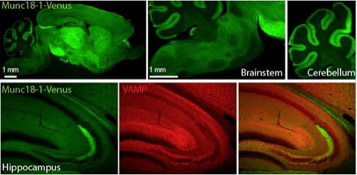
Figure shows expression of Munc18-1-Venus in mouse brain. Munc18-1-Venus is present in almost all brain regions except for specific nuclei in the brainstem.
The lab has previously identified Munc18-1 as essential regulator of vesicle fusion. “This new mouse model opens up a whole new line of research into the function of Munc18-1‚, says Tony Cijsouw. “Previously, we relied on techniques analogous to ‘listening with your ear’ or by studying fixed cells. Now we can look in living neurons what happens with Munc18-1 in synapses before and during activity.‚
“Within the lab, we study mutations in Munc18-1 that lead to severe epilepsy and intellectual disability in patients. These mutations result in reduced expression of Munc18-1. The finding that Munc18-1 is regulated at the synaptic level and that its expression controls synaptic activity is very exciting‚, adds Dr. Ruud Toonen. “Drugs that could influence the synaptic levels of Munc18-1 might help to normalize brain activity in such patients.‚
The paper can be found at The Journal of Cell Biology
