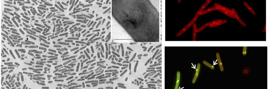
The EM core facility at the VU campus has applied their long-standing experience of monolayer ultrastructural analysis to a different field, the analysis of mircoogranisms such as bacteria.
In collaboration with Michaela Wenzel at the Chalmers University of Technology in Sweden and the team of Lennart Hamoen of the University of Amsterdam, the EM core facility at the VU campus has addressed the ultrastructural effects of antibiotics on the morphology of several bacteria species. Traditionally such studies are done on pelleted cells, which are randomly cross-sectioned for analysis in the transmission electron microscope. This requires large volume cell cultures and involves laborious scanning in EM to find the right cell cross-sections. In the new methodology, a few microliters of cells are seeded on a flat sheet of agar which is then sectioned in parallel. This results in cross sections of hundreds of cells in the same orientation using minimal material. Using this method, we have uncovered that tetracyclin, a well-known antibiotic, causes membrane deformations and relocalization of membrane proteins in B subtilis cells. This effect is independent of the established inhibition of ribosomes by this drug. Using the flat embedding technology, these finding were easily quantifiable over hundreds of cells uncovering effects that previously remained undetectable.
The findings are reported in Communications Biology