Collaboration between CNCR and Max Planck Florida Scientists Identify Neuron Types that Mediate Different Behavioural States
New Findings Provide Insights into the Neural Circuits Responsible for Sensory-Guided Behaviours
In a recent study, scientists from the CNCR & Max Planck Florida Institute have provided one of the most comprehensive analyses to date of the detailed architecture of individual functionally characterized neurons in the cerebral cortex, the largest and most complex area of the brain, whose functions include sensory perception, motor control, and cognition.
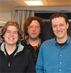
The experiments of this study were performed by Zimbo Boudewijns in the labs of Huibert Mansvelder and Christiaan de Kock at the CNCR and subsequently analyzed using advanced imaging techniques by Marcel Oberlaender in the lab of Nobel laureate Bert Sakmann in Florida. The results of this collaboration were published in the March edition of the Proceedings of the National Academy of Sciences (PNAS). The cutting-edge analyses provide complete three-dimensional reconstructions of the dendritic and axonal anatomy of individual neurons, identify their target neurons throughout the sensory cortical area and describe the information relayed by these neurons during different behavioural states.
Mapping the connectivity within neuronal networks at the level of individual neurons is a major frontier in neuroscience and an essential step towards understanding how the brain works. “Neurons in the brain are grouped into different cell types, each cell type displaying characteristic anatomical and functional properties‚, said Dr. Christiaan de Kock and senior author of the study. “Identifying the three-dimensional pattern of the axon, the neurons’ ‘sending device’, is essential for defining the properties of neural circuits, and, more broadly, establishes the structural constraints that underlie the computational abilities of the brain.‚ The findings from this study will certainly lead to a better understanding of how the cortex transforms sensory information into behavioural responses.

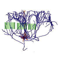
Figure 1: Side view of a reconstruction of a single layer 5 slender tufted neuron from rat vibrissal cortex. Barrel contours are labelled green, dendrites in red/orange and axon in blue. The axonal tree is reconstructed with 1 micron resolution and can reach up to an astonishing length of 94.800 micrometer.