A minimally-invasive brain imaging technique can help researchers visualize individual neurons in live animals without using potentially harmful labels, a study finds.
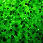
Figure 1: Image of neurons inside brain tissue, as seen by THG microscopy. The neurons are recognizable as dark shadows on a bright background. The actual size of this image is 0.27 × 0.27 millimeters.
Most high-resolution brain imaging techniques for visualizing individual neurons require the use of contrast-enhancing labels, or fluorescent dyes, which could damage neurons or interfere with their function. Stefan Witte and colleagues used an imaging technique called optical third-harmonic generation (THG) microscopy, which circumvents the need for labels, to visualize structures not only in brain slices but also in the brains of anesthetized mice. The technique exploits the specific geometry and lipid content of neurons to create a shadow-contrast image of brain structures in near-real-time. The authors report that THG microscopy enabled rapid, simultaneous imaging of neurons, white matter tracts, and blood vessels without significant damage to brain tissue or changes to neuronal function. Using the technique, the authors visualized individual neurons at depths greater than 300 micrometers in brain tissue and reconstructed the neurons’ approximate shape and location. In anesthetized mice the authors obtained high-contrast images of blood vessels crisscrossing the brain at  depths of about 200 micrometers. The authors suggest that THG microscopy could be potentially useful in brain surgery for precisely guiding microscopic surgical tools and for performing minimally-invasive, real-time, diagnostic brain tissue imaging in people.
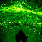
Figure 2: Axon-bundles (white matter) visualized using THG microscopy. These fibrous structures run horizontally through the image. Some neurons can also been seen, as well as a small blood vessel in the lower right part of the image. The actual size of this image is 0.5 × 0.5 millimeters.
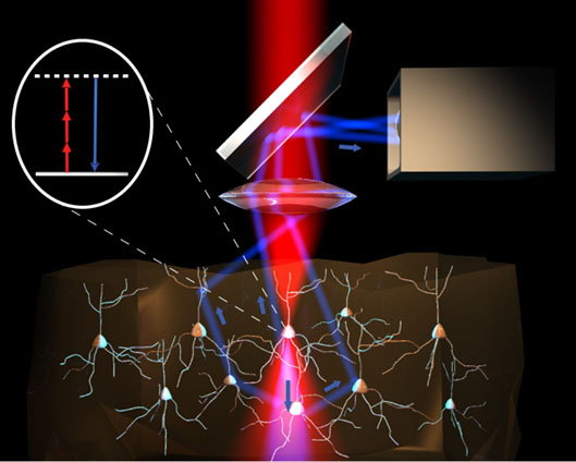
Figure 3: Schematic principle of THG microscopy. Infrared light from a laser is partially converted into blue light (with 3x shorter wavelength) at specific locations, depending on the precise structure of the tissue. By detecting this blue light while moving the laser beam through the tissue, the structure of the tissue can be mapped with high resolution.
Reference
“Label-free live brain imaging and targeted patching with third-harmonic generation microscopy,” by Stefan Witte, Adrian Negrean, Johannes C. Lodder, Christiaan P.J. de Kock, Guilherme Testa Silva, Huibert D. Mansvelder and Marie Louise Groot, Proceedings of the National Academy of Sciences, Published online March 28, 2011.
Contact-informatie:
Stefan Witte
Email: s.m.witte@vu.nl
Tel.: +31-20-5987862
Marloes Groot
Email: m.l.groot@vu.nl
Tel.: +31-20- 5982570
Huib Mansvelder
Email: huibert.mansvelder@cncr.vu.nl
Tel.: +31-20- 5987097
