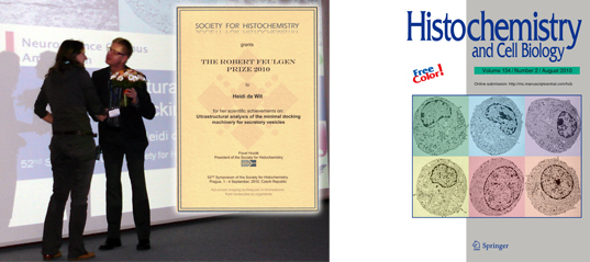
One year after the publication of her Cell paper Heidi de Wit received the Robert Fuelgen Prize 2010.
Exactly one year after the publication of her Cell paper the Robert Fuelgen Prize 2010 was awarded to Dr. Heidi de Wit by the Society for Histochemistry.
Annually, the Robert Feulgen prize is awarded by the Society for Histochemistry for an outstanding achievement in the field of histochemistry either towards the development of new histochemical and cytochemical techniques or in the application of existing technology towards solving important problems in biology and/or medicine (see Feulgen Prize). The award is named after the German physician Robert J. Feulgen (1884-1955) who published many research papers about histochemistry, but he is best known for his discovery of a method for staining nucleic acid, now termed a Feulgen reaction (see also Rober Fuelgen).
On behalf of the Scientific Committee and local organizers, Heidi de Wit was invited to the annual 52nd Symposium of the Society for Histochemistry in Prague, Czech Republic, 1 – 4 September, 2010 as a special invited speaker – the Robert Feulgen Prize 2010 Laureate Lecture. The focus of this Symposium was on ‘Advanced imaging techniques in biomedicine: from molecules to organisms’ (see scientific programme of the 52nd Symposium of the Society for Histochemistry).
During this meeting De Wit received the Robert Fuelgen Prize 2010 for her work on the ‘Ultrastructural analysis of the minimal docking machinery’ from the President of the Society for Histochemistry, Prof. dr. Pavel Hozák (see below).
Heidi de Wit received the Robert Fuelgen Prize (left) from the President of the Society for Histochemistry, Prof. dr. Pavel Hozák (right).
As the Robert Fuelgen Prize 2010 winner Heidi de Wit was also invited to write a review on her awarded work which appeared recently in Histochemistry and Cell Biology the official journal of the Histochemistry Society (see HCB review De Wit 2010). Noteworthy to mention is that her electron microscopic photos were selected for the cover illustration of the issue in which here review was published: Histochem Cell Biol (2010) 134:103–113 (see above).
